TINISCOPE · ULTRALIGHT IN VIVO MICROSCOPY
Multi-region calcium imaging
in freely moving mice
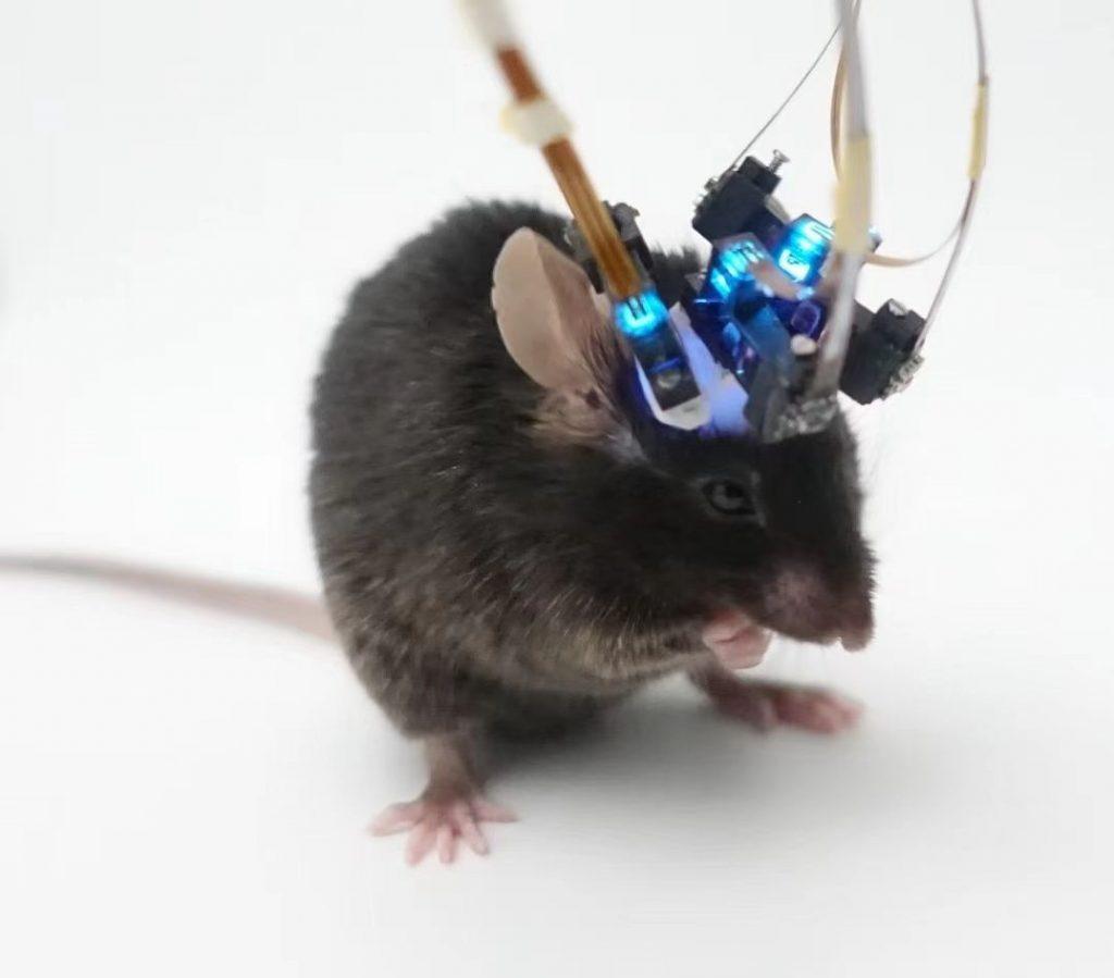
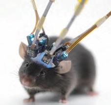
TINISCOPE combines ultralight head-mounted microscopes with synchronized image acquisition and behavioral video. Record calcium signals from neuronal populations in up to four brain regions of the same freely moving mouse and relate activity across regions to behavior.
WHY MULTI-REGION
Relate activity across regions to behavior
Perception, decision-making, memory, and movement involve distributed neural circuits. Simultaneous calcium imaging allows researchers to compare activity across regions within the same behavioral session.
Compare regions within one session
Record from multiple regions in the same animal and session, using a common time reference to compare calcium signal time courses and responses to behavioral events.
Relate calcium signals to behavior
The lightweight head-mounted design supports freely moving experiments, including exploration, learning, and social interaction. Synchronized video helps relate calcium responses to specific behavioral events.
Examine coordinated activity
Compare responses to the same behavioral events and examine shared activity patterns, temporal relationships between calcium signals, and changes across behavioral states.
SYSTEM DESIGN
Compact electronics for a lighter microscope
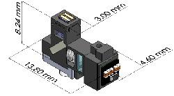
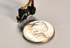
A compact CMOS image sensor with serial output eliminates the need for a separate serializer. Power supply and clock generation are integrated into the data acquisition unit, reducing the size and weight of the head-mounted module while supporting synchronized acquisition from multiple microscopes.
APPLICATION EXAMPLES
Multi-region imaging in practice
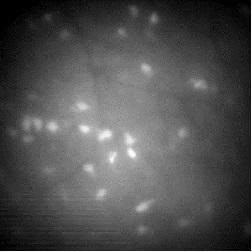
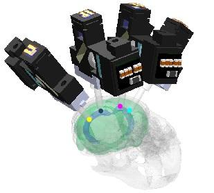
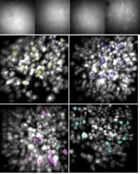
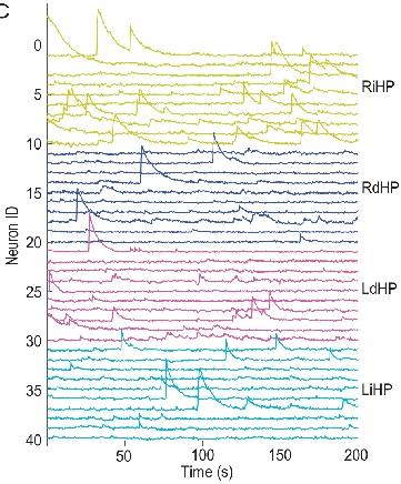
These examples show a four-microscope setup, imaging-site placement, simultaneous recordings from four brain regions, and neuronal calcium signal traces.
SPECIFICATIONS
Technical specifications
The system supports single-photon calcium imaging, simultaneous recording from multiple microscopes, and synchronization with behavioral video. Microscope, cable, and gradient-index (GRIN) lens configurations can be selected to suit the target regions and behavioral task.
- Imaging modality
- Single-photon fluorescence
- Microscope weight
- < 0.5 g
- Objective NA
- 0.52
- Magnification
- 2.67×
- Field of view
- 720 × 540 μm
- Pixel resolution
- 1 μm
- Frame rate
- 25–40 Hz
- Image output
- 10-bit grayscale
- Excitation band
- 470 ± 20 nm
- Emission band
- 525 ± 25 nm
- Microscope dimensions
- 4.6 × 13.8 × 8.24 mm
- Data interface
- USB 3.0 × 2 / × 4
RESEARCH PAPER
The research behind TINISCOPE
Published in National Science Review, the study describes the TINISCOPE design and demonstrates synchronized calcium imaging across multiple brain regions in freely moving mice.